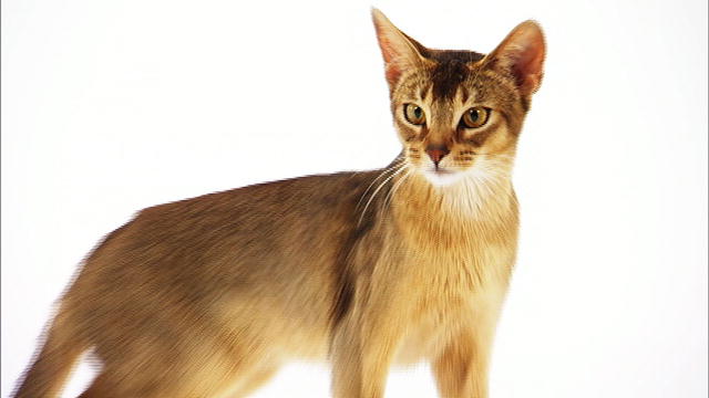
The Breed History
As with many cat breeds, the true origins of this ancient breed have
been lost. One theory is that it was in Abyssinia (now Ethiopia) that
these distinctive cats originated. According to one source though,
genetic studies have indicated their place of origin was in Southeast
Asia and the Indian Ocean coast instead. The first reported export
to Britain was Zula, in 1868. The Abyssinian maintains a distinctive
wildcat look, closely resembling cats depicted in the Egyptian
tombs. By the late 1800s, the Aby, as they are fondly termed, was
well known as a distinct breed. Residual tabby markings are often
visible along the topline, over the eyes, and faint broken bars may
be visible on neck and legs. Note that the breed standard for this
cat varies depending on the registry, with the European type being
more extreme in shape, and a wider spectrum of colors accepted
there. Foundation stock for the American Abyssinian arrived from
Britain in the 1930s. Following WW II, only 12 registered cats
remained in England. The CFA does not allow outcrossing.
Physical Characteristics
Weight: 9-12 lb (4-5.5 kg)
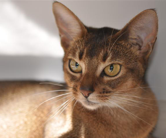
Coat: It is thought that the tabby pattern underlying the agouti
ticked coat was originally much more prominent and that selective
breeding for the ticking and against the tabby resulted in the
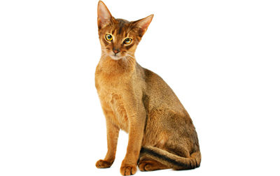
modern coat. Chin and chest are white. The fine, short, shiny, firm
but not harsh hairs have the distinctive agouti pattern. The agouti
typically provides two to four color bands over the shaft of the hair,
with the ticking color at the terminus of the hair and the base of
the hair being the main coat color. Names below that are bolded are
accepted in CFA. Others are accepted in European registries. Agouti
is a dominant coat factor (A).
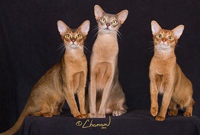
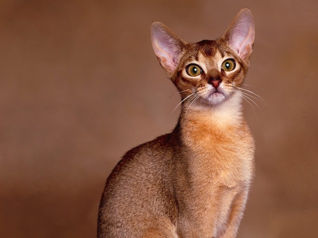
Ruddy: The most popular color, the "Usual" Abyssinian coloring is
a rich medium honey brown (burnt sienna) ticked with dark brown/
black.
Blue: Is a base coat of pale beige (oatmeal) with ticking of
bluish-grey (slate blue).
Sorrel: The so-called red color is popular-it's a rich ginger red (also
termed apricot); ticking is a dark rich chocolate brown. This is not a
true red (red is a sex-linked recessive).
Fawn: A pale orange (rose beige) base color with warm milk
chocolate (cocoa) ticking.
Lilac and silver: Not yet accepted in North America.
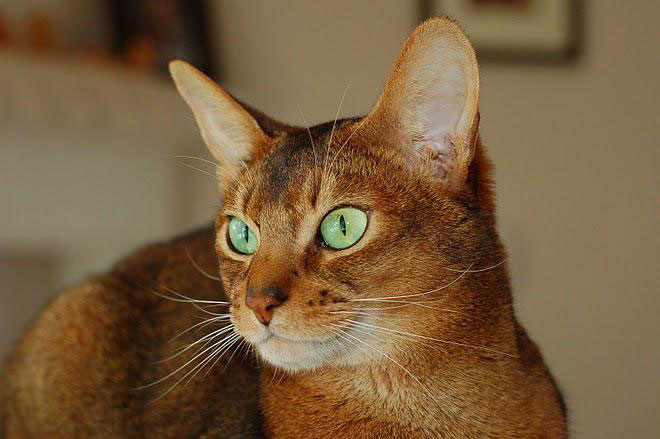
Eyes: The large almond eyes are fairly wide set; may be green or
gold in the CFA-approved colors. There are dark rims set within a
lighter spectacle.
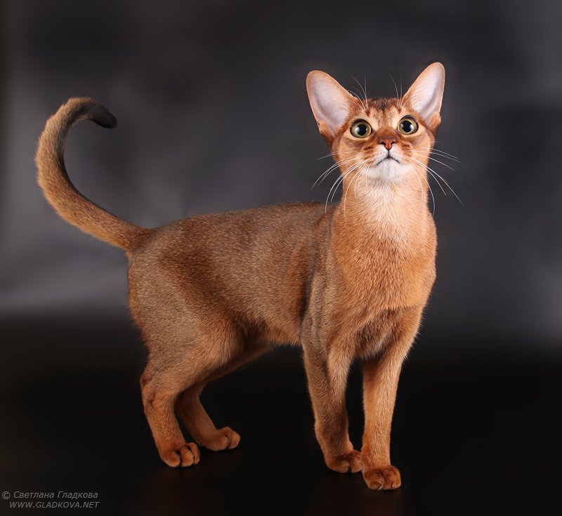
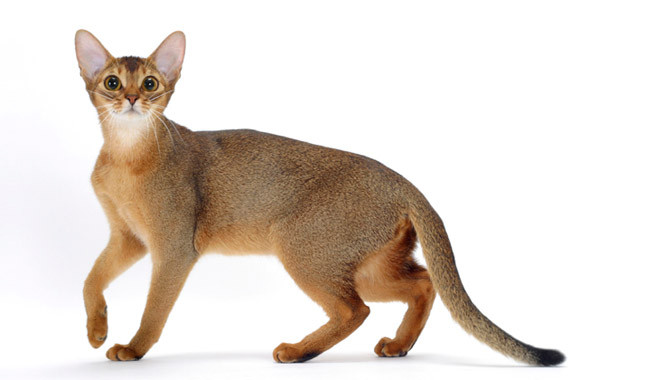
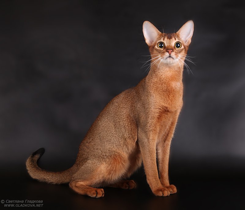
Points of Conformation: The Abby is a lithe, medium-sized cat
with slender legs and long arched neck. The wide set moderately
sized ears are tipped forward. The face and head has a distinct
rounded-wedge shape, without flat planes. The English Abyssinian
has a more elongated head when compared with the American cat.
There is a slight break of the nose. Feet are small, compact, and
oval. Chin hairs are light. Tail terminus is the ticking color and the
tail is the same length as the body; fine and tapering from a thick
base. Back is slightly arched.
Grooming: Low grooming requirements. A chamois cloth or hand
grooming once weekly will usually suffice.
Recognized Behavior Issues and Traits
Reported breed characteristics include: Athletic, playful, very active,
affectionate, curious, highly intelligent, independent, love to jump
and be in high places. A few lines have high strung/nervous or
independent temperament. They tend to become very attached to
their caregivers and may demand attention and shadow. Highly
social, they enjoy the company of other cats in the household;
a busy-body. Quiet voiced cats. Play fetch and ride shoulders-
recommended to have climbing trees. Many like water and will use
their paws to drink and play in water.
Normal Breed Variations
Small litters
Quick to mature
Drug Sensitivities
None reported in the literature except anecdotal reports of
sensitivity to griseofulvin.
Inherited Diseases
Systemic AA/Renal Amyloidosis: Amyloidosis can occur in chronic
inflammatory diseases (secondary), or be inherited (primary).
Amyloidosis is considered a primary condition in this breed
(and the Somali). Glomerular and medullary interstitial amyloid
(amyloid AA, an inert beta pleated fibrillar protein) deposits, with
papillary necrosis and medullary interstitial fibrosis resulting. Renal
amyloidosis is considered a subset of systemic amyloidosis; in most
if not all cats, deposits occur in many organs (e.g., thyroid) but the
primary clinical picture results from the renal deposits. Resulting
chronic renal failure is typical in clinical presentation and occurs in
young cats. Severe proteinuria is uncommon in the cat (compared
with amyloidosis in dogs) because the most severe changes affect
the medullary interstitium (rather than the glomeruli, as is the case
in canines).
Typical age of presentation is 1-5 years old; the female to male ratio
is 1.4:1, and first evidence of renal amyloid deposits in the kidney
can be found at 9-24 months of age. An autosomal dominance
inheritance with variable penetrance was proposed. In some cats,
by one year later renal failure may be evident while in others,
deposition of amyloid is slow and they may live a clinically normal
full lifespan. A surgical biopsy of both renal medulla and cortex is
most likely to be diagnostic. Apart from routine H&E, a Congo red
stain is performed to highlight amyloid.
In a summary of generalized AA amyloidosis in various cat breeds
(especially Siamese and Oriental cats 1987-1994), amyloidosis was
reported to be a familial trait in the Abyssinian.
In a study of cats (1983-1997; Netherlands), 3.1% of 258
Abyssinian referrals were diagnosed with AA-amyloidosis. Affected
cats were quite inbred. In 25% of the Abyssinian cats with
amyloidosis, inflammatory conditions were present concurrently
(e.g., rhinitis, FIP).
The amyloidosis [apo-lipoprotein-serum amyloid A (AA)] propensity
is still not clearly elucidated as a solely genetic problem; sequencing
of the proteins shows amino acid shifts common to different breeds
and this may mean that in addition to the presence of amyloid
associated genes, other factors such as chronic inflammation or
certain infections are involved in the genesis of phenotypic clinically
significant amyloidosis. There may be three genes involved.
Recessive Hereditary Rod-Cone Degeneration rdAc/Progressive
Retinal Atrophy [PRA]: Age of onset is usually early adulthood
(1.5-2 y), with slow progression to generalized retinal atrophy by
middle age. A plasma lipid abnormality was found in conjunction
with the eye problem-a reduced plasma level of docosahexanoic
acid, an omega-3 fatty acid was documented. This fatty acid is the
major fatty acid in outer segments of retina rods. This condition is
similar to retinitis pigmentosa in humans.
It was determined that an autosomal recessive pattern of
inheritance was occurring. In an early report of Swedish cats,
indirect binocular ophthalmoscopic examination proved that of
205 cats assessed over 2 years old, 68 had bilateral disease (34%),
while 45% were affected in one or both eyes. At 1.5-2 years of
age, the first ophthalmoscopic changes were noted, and peripheral
tapetal fundus was discolored brown to grey, along with retinal
vessel attenuation. By about three years of age, tapetum color had
changed to gray. Late stage, there was tapetal hyper-reflectivity,
and severe vessel attenuation was noted; PLR was reduced or
absent. Latest age of onset was at four years of age.
It was reported that when homozygous for the trait, affected
kittens had an abnormal ERG as early as eight weeks of age; the
cat retina is considered fully developed at about 10 weeks of age.
Ophthalmoscopic examination in those cats can remain normal
until almost two years of age. This study also confirmed an early
drop in the levels of inter-photoreceptor retinoid-binding protein
(IRBP) starting at four to six weeks of age in homozygous kittens.
This protein also binds fatty acids at the retina.
The even distribution of retinal pathology during early phases changes
to retention of function preferentially in the central retina, with loss
of function peripherally in later stage disease. Retina blood flow was
found to be severely compromised in a study of cats with late stage
PRA. Antibodies against both green- and blue-sensitive cones were
used to establish that early reduction of both of these cone types
occurs, while the inner retina is mostly preserved. Late in disease, the
ERG is not recordable and the retina is markedly thinned.
Recent data using electron microscopy proves that the arterial walls
of vessels in the iris are abnormal even though innervation is normal.
The ciliary processes were also shorter and more compact than in
normal cats.
A closed colony was studied to further elucidate the correlation with
phenotype and genotype. The rdAc allele was found in European and
Australian cats with moderate frequency.
Dominant Abyssinian Rod-cone Dysplasia (Rdy): A second type
of retinal condition has been described in Abyssinians which has
an autosomal dominant mode of inheritance. Visual deficits are
congenital, and horizontal nystagmus may also be noted. At four
weeks of age mydriasis is noted, and significant visual deficits or
blindness occur by 12-16 weeks of age. By two weeks of age, early
retinal changes are occurring and the rods and cones are equally
affected. Progressive loss occurs from central retina towards the
periphery. This condition has only been reported in the UK. This is a
true dysplasia as rods and cones never fully develop.
A single mutation was identified as a single base pair deletion on
the Exon 4 of the CRX peptide gene in the cat, which interferes with
formation of the key protein.
Arterial Thromboembolism: In one study of 127 affected cats,
Abyssinian cats were one of three breeds overrepresented with
arterial thromboembolism.
Blood Type B: There may be geographic variation since in a small
study of cats in Hungary, none of the cats tested were of B blood
type.
Prevalence of B blood type in the USA was 20% in one report.19 In
another study of blood type distribution in the USA, 230 Abyssinian
cats had a prevalence of 13.5% blood type B, and in a summary of
studies 16% pooled prevalence of type B was reported.
In Australia, all PK cats tested (n=36) were determined to be type A.
(See below for more about PK deficiency).
Neonatal Isoerythrolysis (NI): The reported proportion of matings
at risk for NI is 0.12.
Pyruvate Kinase Deficiency and Increased Osmotic Fragility of
RBCs: The pyruvate kinase enzyme (PK) is involved in the anaerobic
glycolytic pathway of erythrocytes. Lack of the enzyme function
results in energy depletion, and premature red cell destruction. Cats
with this condition may experience a range of severity of anemia,
with some experiencing recurrent severe hemolytic anemia and
splenomegaly.
In one study, osmotic fragility occurred in the absence of PK
deficiency. Reported onset of anemia ranged from 6 months to
5 years of age (mean 23 months) and typical PCV ranged from 15%-25% (as low as 5%). Hepatic enzymes were elevated in some
of the cats. Macrocytic anemia with reticulocytosis occurred.
Macrocytosis persisted when the anemia resolved. Osmotic fragility
tests indicated much higher fragility than normal. Some of the 18
affected cats with this primary erythrocyte fragility were closely
related (both Somali, Abyssinian) and an autosomal recessive mode
of inheritance was postulated.
In another study of Abyssinian and Somali cats aged 1-10 years,
chronic intermittent hypochromic regenerative hemolytic anemia and
mild splenomegaly were reported. Pyruvate kinase activity ranged
from 6%-20% of normal. Since parents were less severely affected,
it was postulated to be an autosomal recessive condition-now
confirmed. Osmotic fragility was normal to mildly increased.
In a case report, a one year old male Abby cat presented with
splenomegaly, mild exercise intolerance and severe regenerative
hemolytic anemia (Coomb's negative). Splenectomy produced
partial remission at 1.5 years of age; proband PK enzyme activity
was 15% of normal and the proband queen's PK enzyme activity
was 50% of normal.
Affected Somali and Abyssinian cats usually have a normal lifespan,
unlike dogs with this condition (see Beagles, Basenji, Westie,
Dachshund). Dogs tend to develop liver failure and osteosclerosis
while cats do not. Enzyme analysis and molecular genetic testing are
available at the Deubler Laboratory-see below under Genetic Testing.
Anemia may be noted in cats as young as six months, and has been
found in senior cats (12 yrs old) that were clinically normal.
A recent study showed not all cats between 1-11 years of age
developed clinical signs, though a mortality rate of about 25%
(presumed to be due to PK) occurred in the study group. In a little
over half of the cats (median age 1.7 y), lethargy, diarrhea, poor
appetite, and weight loss, pale mucous membranes and icterus were
most commonly noted. Symptomatic cats had variable lab findings
with increased bilirubin, globulins, liver enzymes and reticulocytes.
The authors say "As PK-deficient cats can be asymptomatic testing
for PK deficiency before breeding is strongly recommended."
Disease Predispositions
Renal Failure: Renal failure rate for Abyssinian cats was identified
to be more than double the baseline rate in a study of cats seen
at veterinary colleges between 1980-1990 (189,371 cases in the
Purdue University Veterinary Medical Database), at an odds ratio for
risk of 2.42:1 (22 of 771 cats, prevalence 2.85%).
Cervical Neck Lesions: Increased risk was reported. Abyssinians
were one of two breeds (the other being Siamese) most commonly
affected by feline odontoclastic resorptive lesions (FORLs).
Gingivitis: Anecdotal evidence for increased prevalence.
Psychogenic Alopecia, Hyperesthesia: Anecdotal reports of
increased prevalence.
Urinary Tract Infection (UTI): In a study of 22,908 cats with feline
lower urinary tract disorders, Abyssinian cats were overrepresented
for UTI.
Feline Dilated Cardiomyopathy (FDC): Breed disposition was
reported for FDC; the hypertrophic form is rarely reported in this
breed.31 Average age of onset is 7 years. Case rates have dropped
since diet supplementation with taurine began but is still seen.
Previously, mortality for FDC was about 85%, but with taurine
supplementation mortality is currently in the 30%-50% range.
Cats don't typically cough as in the dog; heart rate may range from
bradycardic to tachycardia, and both ventricles are affected. Pleural
effusion, cold extremities, weakness and azotemia are typical.
Affected cats are prone to arterial thromboembolism. Systolic
heart murmur or diastolic gallop can often be heard, and 61%
have arrhythmias, usually ventricular. Echocardiography is the best
modality for definitive diagnosis.
Medial Patellar Luxation (MPL)/Hip Dysplasia (HD): In one study
it was noted that of 69 Abyssinian cats, 26 (38%) had abnormal
patella seating with easily induced luxation; a possible dominant
(but polygenic) inheritance was suggested. Minimum age was 6
months; average age of the study group of cats was 4.3 years.
This study group represented several US and European lines in the
breed; diagnosis was by manual palpation to displace the patella.
In a mixed breed study looking at medial patellar luxation and
hip dysplasia at the University of Pennsylvania (1998) of 78
non-randomly selected cats over six months of age (average age
2.5 yr), Abyssinians were afflicted with MPL more frequently and
severely than the average cat. Cats with MPL in the pooled group
(including the Abyssinian) were three times as likely to have HD
as those without MPL. Only 11 of the 78 cats had clinical signs of
pelvic limb abnormalities so many cats are not being diagnosed in
clinical practices. Norberg angle (NA) and distraction index (DI) in
combination with OFA criterion were used to assess the cats. They
found 80% of Abyssinian cats (8/10) had MPL and of these, 7/8
had bilateral MPL. And 33% (3/10) had HD, while 33% (3/10) had
concurrent HD/MPL. A positive correlation between joint laxity and
HD was identified.
FIP Susceptibility: An American Study found that Abyssinian cats
were significantly over-represented for a diagnosis of FIP when
they analyzed data for a 16 year period at a veterinary teaching
hospital.
Rare and Isolated Reports
Congenital Hypothyroidism: Primary dyshormonogenesis has
been diagnosed in a group of related Abyssinians that had early
onset disproportionate dwarfism, goiter, constipation and retained
kitten features. It was reported to be inherited as an autosomal
recessive disorder. This trait results in thyroid peroxidase deficiency.
By four weeks of age kittens were stunted, by six to nine months
of age still immature, and by adulthood, variable expression was
noted. Deciduous teeth were shed late and epiphyseal plate closure
was delayed. Both TSH and TRH tests were abnormal, and the t-T4
and T3, and f-T4 low. This was proposed to be an under-diagnosed
cause of fading kittens. If detected early and treated with
appropriate thyroid hormone supplementation, effects may be
reduced or ameliorated.
Myasthenia Gravis (MG) and Thymoma-associated
Neuromuscular Disorder: Can be a congenital (Siamese) or
acquired condition (Aby, Somali). One study reported that compared
with mixed-breed cats, relative risk for acquired MG is highest
in Abyssinian (and Somali) cats; relative risk also increased
after three years of age. The MG is associated with dysphagia,
megaesophagus, weakness, or weakness with a cranial mediastinal
mass.41 Acetylcholine receptor antibodies produced within/by the
thymoma and in muscle cross reacted. Clinically, voice changes,
tremor, ventroflexion of the neck and gait abnormalities were
noted. Myasthenia gravis signs are typical of the acquired idiopathic
form. Acetylcholine receptor antibody titers of > 0.3 nmol/L are
diagnostic for acquired MG. Some afflicted cats were noted to have
been treated for hyperthyroidism with methimazole for 2-4 months
prior to the diagnosis of MG in one report. The study did not
attribute any causative effect though.
Mycobacterium avium Susceptiblity: In North America and
Australia, of a case series of 12 cats under 6 years old, 10/12
were Abyssinian, and 1/12 was Somali, a closely related breed.
Clinical signs include weight loss, lower respiratory tract infection,
and enlarged lymph nodes, with slow re-growing hair. A familial
immune-compromise was suspected in certain lines. They were
FeLV-FIV negative cats but developed disseminated infection.
Long term treatment with clarithromycin combined with another
agent such as rifampicin and a fluoroquinolone, or doxycycline was
somewhat effective. It was proposed that there are certain lines of
Somali and Abyssinian cats that are predisposed due to a familial
immunodeficiency.
Glycogen Storage Disease: A 10-year old cat in Indiana was
diagnosed with a storage disease that resulted in accumulation of
what appeared to be unbranched glycogen in skeletal muscle, and
minor deposits were in cardiac muscles, brain and spinal cord. Onset
of signs occurred gradually, with paresis progressing to paralysis.
Genetic Tests
Progressive Retinal Atrophy: Direct tests for rdAc and Rdy are
available at the UC-Davis VGL.
Pyruvate Kinase Deficiency DNA Test: This is a direct test (not
linkage) which identifies affected, carrier, and normal cats. Testing
is recommended for anemic Abyssinian and Somali cats, relatives of
affected or carrier animals and breeding stock unless all parents of
the breeding pair have previously tested negative. Check with the
local lab but generally, a minimum of 1-2 ml EDTA blood should be
sent immediately (not frozen-cooling the sample is not essential).
It should be processed within 48 hours. Test request form available
online. A pair of buccal swabs can be used, but one must ensure
cells are sampled, not just saliva. Concurrent blood typing can be
carried out on the same blood sample. Available at PennGen and
UC-Davis VGL.
Renal Function: Screen renal function (as a minimum, serum
creatinine and urinalysis) in cats eight years and older since a breed
propensity to renal failure has been identified.
Electroretinogram (ERG): Cats with inherited rod-cone
degeneration can be screened using ERG in the pre-clinical phase to
identify cats at risk for progression. A study comparing homozygous
affected and heterozygous at risk cats confirmed that cats with the
inherited condition will show the following changes:
Decreased a-wave amplitude
Increased b-wave implicit times
Abnormal ERG waveforms
b-wave:a-wave ratios high
A graphic representation of results can distinguish between
genotypes, even before funduscopic changes are evident.
Another study showed that short ERG protocol is more efficient
than a long one in the identification of affected cats. A study using
12 parameters was adequate to discriminate rod-cone dystrophic
cats from normal cats.
Anti-acetylcholine receptor antibodies (serum) for MG: Send to
Comparative Neuromuscular Laboratory, att: Diane Shelton, UCSC
http://vetneuromuscular.ucsd.edu/
No genetic tests are yet available for rdAc or Rdy eye diseases.
Miscellaneous
- Breed name synonyms: Aby, Ticked Cat, Egyptian Cat, informal
name "Bunny Cat"
- Registries: FIFe, TICA, CFA, ACFA, CFF, CCA, NZCF, WCF, ACF, GCCF
- Breed resources: CFA Abyssinian Breed Council:
www.abyssinianbc.org
The Abyssinian Cat Club of America: http://www.abyworld.com/
Abyssinian Cat Association (UK): www.theabycat.com
Photo Gallery of Breed - Abyssinian - Cat Breed
 Animalia Life